第1回テーマ:形状測定

午前の講義と午後の実習に分け、じっくりと装置と向き合い、正確なオペレーションを身に着けていただくことが目的です。
これからオペレーションを始められる方、普段の測定で疑問点がある方、測定方法について相談のある方にオススメです。
〇対象 :Park装置をお使いのユーザー様
〇開催日時:2023年5月18日(木)10:00~17:00
2023年5月19日(金)10:00~17:00
※18日と19日は同じ内容となります。どちらか1日を選択いただきお申込ください。
〇プログラム:
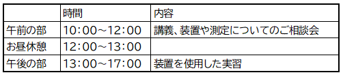
午前中は、講義を1時間、測定やプローブ、装置のアップグレードや今後の運用についてのご相談会として1時間設けております。
午前中のみのご参加もぜひお気軽にお申込みください。
〇開催場所 :パーク・システムズ・ジャパン株式会社 デモルーム
〒101-0054 東京都千代田区神田錦町1-17-1 神田髙木ビル1階
東京メトロ半蔵門線 竹橋 3b出口 徒歩6分
JR 中央線 山手線 京浜東北線 神田 西口 徒歩9分
〇開催方法 :現地開催
〇申込方法 :以下フォームよりお申込みください。
実習は定員を設けております。(定員:6名)満席になる可能性がございますのでご了承ください。
〇お問い合わせ先:パーク・システムズ・ジャパン株式会社 担当:徳升
☎:03-3219-1001 ✉:[email protected]
Ferroelectricity is observed in hexagonal boron nitride(hBN) through control of the registry of stacked layers, which we explore through both amplitude-modulated and sideband Kelvin probe force microscopy (KPFM) on the Park FX40 automatic AFM.
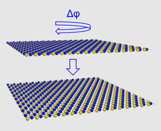
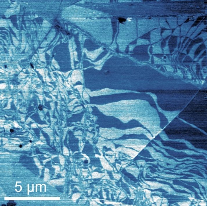
A schematic of the formation of parallel stacked bilayer hBN is shown in addition to a contact potential difference map measured using sideband KPFM.
Image caption
Image caption
Image caption

James received his PhD in Physics from the University of Nottingham in 2018, studying the morphology and optical properties of monolayers of self-assembled molecules and their heterostructures. He then went on to work as a postdoctoral researcher, also at the University of Nottingham, working on the formation of hybrid heterostructures of molecular assemblies and layered materials demonstrating both electroluminescence and selective triplet excitation. In 2020, James took up a position as a postdoctoral researcher at the Cambridge Graphene Centre, using scanning probe microscopy and optical spectroscopy to study electrostatics and optical properties of layered materials heterostructures with controlled twist angle and their scalable incorporation into integrated photonic circuits. Since January 2022, James has been a member of the Park Systems team as an applications scientist, supporting customers with interest ranging from fundamental physics to industrial scale production in the application of a diverse range of scanning probe microscopy techniques to gain insightful results.