Webinar Series | April 2023

Giovanni Longo, Simone Dinarelli, Marco Girasole
Istituto di Struttura della Materia – Consiglio Nazionale delle Ricerche
Atomic force microscopy (AFM) is an extremely versatile technique enabling the study of different aspects of biological systems with ultra-high resolution. Conventionally, AFM has been used to assess the morphology of living biological systems and to monitor the evolution of their mechanical properties at the nanoscale, which provides information on the cellular structure and stiffness even in living systems. Further applications of AFM for biological matter involve the combination of ultrastructural information with conventional or fluorescence optical microscopy. Correlative analyses via combining advanced AFM with high-magnification optical and fluorescence microscopes allow for a more comprehensive understanding of the cellular status.
In this webinar, we will use the Park Systems NX12 AFM coupled with a Nikon fluorescence microscope to perform studies on living biological systems such as erythrocytes and neuroblastomas. By using conventional optical and fluorescence images, we identify cells that are most interesting for our studies. We use the AFM to determine their morphology and mechanical properties at ultra-resolution level, monitoring the response of the cells to environmental conditions such as pharmacological stimuli or starvation. Also, we prove how AFM cantilevers can be used as nanomechanical sensors to provide complementary information on the cellular behavior in different environments.
The combined results on neuroblastomas represent the first steps of the COMA-SAN project (COMplexity Analysis in the Simplest Alive Neuronal network), in which we investigate the communication-mediated group behavior of these cells. Overall, our study opens a path to better understand the interactions between cells and to evidence the complexity of group dynamics in cells.

Figure 1. Correlative Optical imaging/AFM morphology on favism red blood cells

Figure 2. Evolution of living neuroblastomas exposed to pharmaceutical stimulation

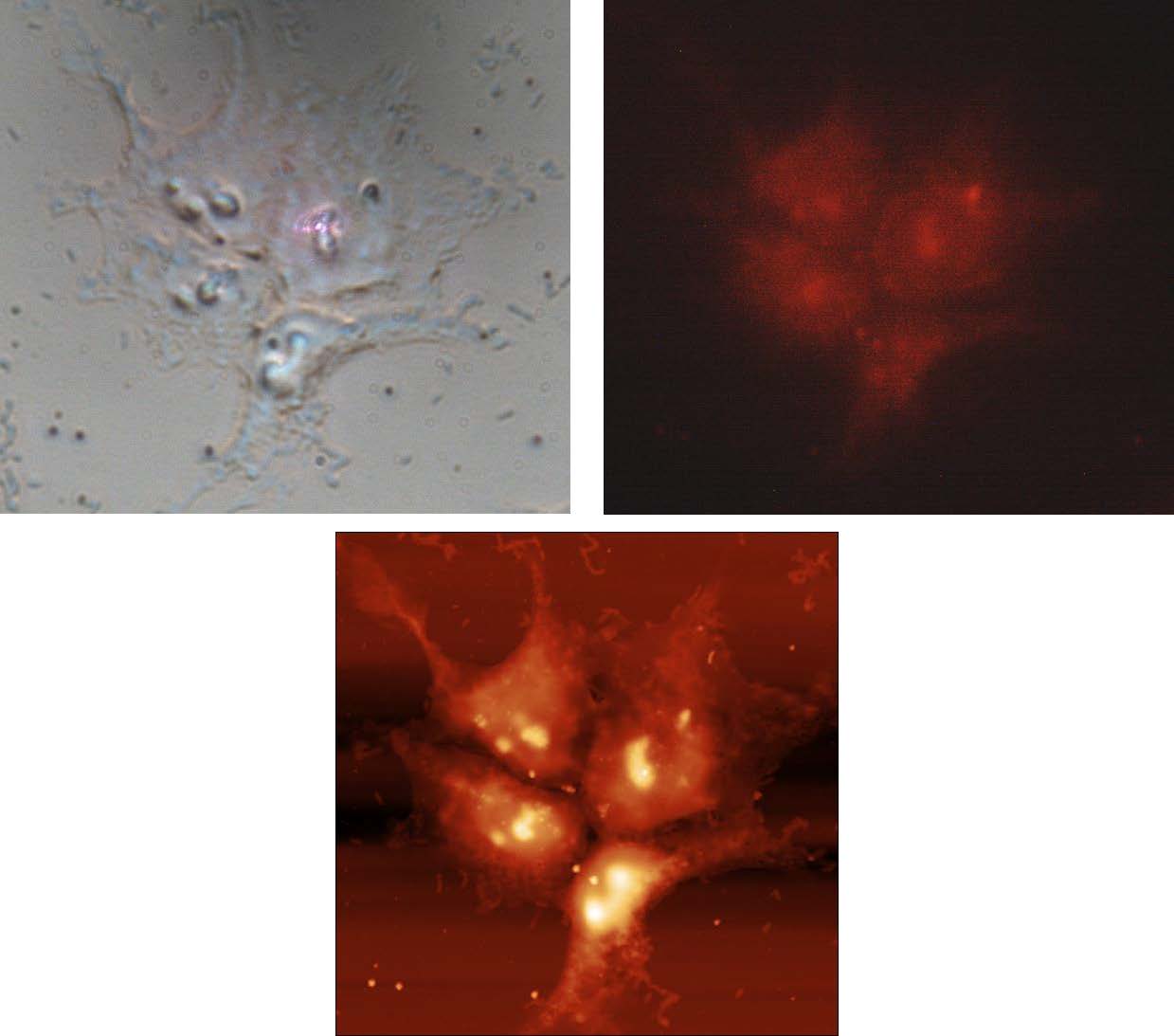
Figure 3. Correlative Optical imaging/Fluorescence imaging/AFM morphology on HeLa cells
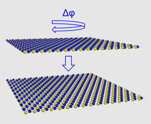
Fig. 1. A schematic of parallel stacked 2L-MoS2 on HOPG (a) is shown


Image caption

Image caption

Dr. Giovanni Longo is a researcher at the Consiglio Nazionale delle Ricerche in Rome, at the Istituto di Struttura della Materia. He was awarded his Ph.D. in Materials Science from the University of Rome La Sapienza in 2006. From 2010 to 2015 he worked as a senior Postdoc at the EPFL in Lausanne in the LPMV laboratories. He is currently head of the BioLab in the ISM-CNR and in the organizing committee of the Tech4Bio consortium, aiming to further the interactions between biophysics research and industries. As an expert in scanning probe microscopies and high-resolution characterization techniques, he has focused on the interdisciplinary studies combining physics and biology for biomedical applications, employing AFM in innovative ways, and combining this technique with Raman or IR spectroscopy. Expanding on this background, he is among the main developers of the Nanomotion sensor, which he has applied to different experimental fields. At the same time, he and his group are working on several scientific topics, ranging from the study of the presence of nano-pollutants in living systems, to the effect of mechanical stimuli on mammalian cells, including Red Blood Cells and neurons. Currently he is investigating the collective movements of groups of living cells (i.e. neurons), to determine external stimuli and the group size can cause the rise of emerging properties.
